


Joints and Movement
Types of joints
- Joints form where two or more bones come together
- There are three main types of joints:
- Fibrous:
- Held together by fibrous connective tissue
- No joint cavity between the bones
- Immovable
- E.g. between the various bones of the skull
- Cartilaginous:
- Connected together by cartilage
- Allow partial movement – a bit more movement than fibrous joints but much less than synovial joints
- No joint cavity between the bones
- Further subdivided into:
- Synchondrosis (primary cartilaginous joint):
- Bones are joined by hyaline cartilage
- Most of them are the growth plates and are therefore only present in children until puberty
- once growth stops the growth plates become calcified and so fuse to become solid bone
- An example of synchondrosis that remains into adulthood is that between the first rib and the manubrium sternum
- Symphysis (secondary cartilaginous joint):
- Bones are joined by fibrocartilage
- Located in the midline plane of the skeleton
- E.g. pubic symphysis located at the front of the pelvis has fibrocartilage
- E.g. intervertebral discs located between the vertebrae of the spine
- Synovial:
- They are the most abundant type of joint in the body
- Characterised by three main structures:
- Synovial cavity:
- The enclosed space around the joint surfaces is filled with synovial fluid which is the lubricant of the joint
- Articular capsule:
- Composed of fibrous connective tissue and forms the wall of the joint
- The inner surface of the articular capsule is lined by synovial membrane
- The cells of the synovial membrane produce the synovial fluid
- Articular cartilage:
- The ends of the articulating bones are lined by articular cartilage which helps reduce friction to movement
- Unlike cartilaginous joints, the cartilage on each bone is not continuous with each other and instead allow movement between each other
- There are 6 different types of synovial joints:
- Ball and socket joint:
- They comprise of a spherical end of a bone articulating with a dished end of another bone
- Due to their shape they allow the maximum range of movement: flexion, extension, abduction, adduction, rotation, circumduction
- E.g. shoulder and hip joints
- Hinge joint:
- Like a hinge of a door, the majority of movement allowed in these joints is in one plane: flexion and extension
- E.g. elbow; the knee is primarily a hinge joint but is more complex as it also allows some rotation and translation
- Saddle joint:
- Shaped like a saddle of a horse
- Similar to hinge joint but oval shape of the saddle provides greater range of motion
- Movements allowed: flexion, extension, adduction, abduction and circumduction
- They do not allow axial rotation
- E.g. at the base of the thumb (called first carpometacarpal joint)
- Condyloid (ellipsoid) joint:
- Shallow depression of one bone articulates with rounded end of the other bone
- Movements allowed: flexion, extension, abduction, adduction, circumduction
- They do not allow axial rotation
- E.g. metacarpophalangeal joints of the hand and metatarsophalangeal joints of the foot
- Pivot joint:
- They only allow axial rotational movement
- E.g. the top two vertebral bones in the neck (atlas and axis)
- Gliding (plane) joint:
- Allow only for sliding movement between small bones with flat surfaces in the plane of the articular surface
- E.g. small bones in the wrist and foot


Functional classification of joints
- Joints can also be classified according to the degree of movement they allow:
- Synarthrosis:
- These are the fibrous joints
- Permit little to no movement
- Amphiarthrosis:
- These are the cartilaginous joints
- Permit little mobility
- Diarthrosis:
- These are the synovial joints
- Allow the greatest amount of mobility
Anatomical Terminology
Directional terms
- Superior:
- Towards the head end of the body
- E.g. the head is superior to the hip
- Inferior:
- Towards the foot end of the body
- E.g. the foot is inferior to the chest
- Anterior:
- Towards the front
- E.g. the patella sits on the anterior aspect of the knee joint
- Posterior:
- Towards the back
- E.g. the ears lie posterior to the nose
- Medial:
- Towards the midline of the body
- E.g. the nose is medial to the ear
- Lateral:
- Away from the midline and towards the periphery of the body
- E.g. the ear is lateral to the nose
- Proximal:
- Towards the point of origin or trunk of the body
- E.g. the hip is proximal to the knee
- Distal:
- Away from the point of origin or trunk of the body
- E.g. the ankle is distal to the knee
- Superficial:
- Towards the surface of the body
- E.g. skin is superficial to a muscle
- Deep:
- Away from the surface of the body
- E.g. muscle is deep to the skin
- Ipsilateral:
- Same side of the body
- E.g. the right hand is ipsilateral to the right knee
- Contralateral:
- Opposite side of the body
- E.g. the left knee is contralateral to the right knee
- Unilateral:
- One side of the body
- E.g. Lifting a weight with just one hand is a unilateral exercise
- Bilateral:
- Both sides of the body
- E.g. Lifting a weight with both hands is a bilateral exercise
- Varus:
- Distal part is closer to the midline than the proximal part
- E.g. genu varum is when the foot is closer to the midline than the knee also known as bow legged as the lower limbs take on the shape of a bow
- Valgus:
- Distal part is further away from the midline than the proximal part
- E.g. genu valgum is when the foot is further away from the midline than the knee also known as knock kneed as the knees are knocking (touching) against each other
Planes of the body
- Coronal (frontal) plane:
- A vertical plane that runs from side to side
- E.g. looking at someone from the font
- Sagittal (lateral) plane:
- A vertical plane that runs from front to back
- E.g. looking at someone from the side
- Axial (transverse) plane:
- A horizontal plane that is 90 degrees to the length of the body
- E.g. looking at someone from above
Description of movement
- Flexion and extension occur in the sagittal plane:
- Flexion (bending):
- Reduces the angle between the body parts
- E.g. flexion of the elbow brings the hand towards the shoulder
- E.g. flexion of the knee brings the foot towards the buttocks
- Hyperflexion is excessive flexion of a joint
- Extension (straightening):
- Increased the angle between the body parts
- E.g. extension of the elbow brings the hand away from the shoulder
- E.g. extension of the knee brings the foot away from the buttocks
- Hyperextension is excessive extension of a joint
- Abduction and adduction occur in the coronal plane:
- Abduction:
- Moves the limb away from the body
- E.g. abduction of the shoulder is movement of the arm laterally away from the body
- E.g. spreading of the fingers or toes apart, away from each other
- Adduction:
- Moves the limb towards the body
- E.g. adduction of the shoulder is movement of the arm medially towards the body
- E.g. bringing the fingers or toes towards each other and closing the gap between each finger or toe
- External and internal rotation:
- External (lateral) rotation:
- Rotation of the limb so that the anterior surface rotates away from the midline
- E.g. external rotation of the hip moves the anterior thigh to face away from the midline whilst the sole of the foot faces towards the midline
- Internal (medial) rotation:
- Rotation of the limb so that the anterior surface of the limb rotates towards the midline
- E.g. internal rotation of the hip moves the anterior thigh to face towards the midline whilst the sole of the foot faces away from the midline
- Circumduction:
- Movement in a circular manner
- E.g. circumduction at the shoulder joint involves moving the arm in a circle
- Supination and pronation:
- Rotational movement at the forearm or foot
- Supination brings the palm or sole of the foot facing forward
- Pronation brings the palm or sole of the foot facing backwards
More relevant information can be found on:
Introduction De Quervain’s tenosynovitis (also called De Quervain’s tendinosis) is a painful condition of two tendons at the level of the wrist on the side of the thumb It’s a repetitive strain injury due to overuse of two tendons used to move the thumb away from the other fingers
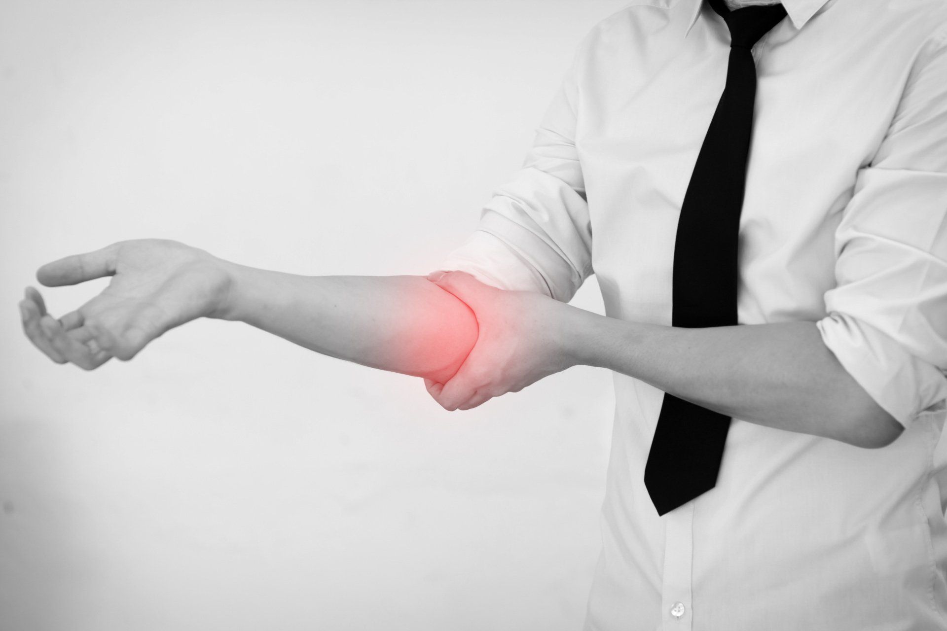
Introduction Cubital Tunnel Syndrome occurs when the ulnar nerve is compressed within a tunnel on the inner (medial) side of the elbow just behind the bony prominence of the inner aspect of the elbow called the medial epicondyle Cubital Tunnel Syndrome is the second most common cause of peripheral nerve compression: The most common one being carpal tunnel syndrome (compression of the median nerve at the wrist) The ulnar nerve is one of the three main nerves of the upper limb: The other two nerves of the upper limb are the median nerve and the radial nerve The ulnar nerve travels from the neck past the elbow and wrist and into the hand: Along the way it travels past some narrow areas where it can be constricted and cause symptoms for the patient The most common site of ulnar nerve compression is in the cubital tunnel at the elbow The second most common site is in Guyon’s canal in the hand When someone accidentally hits the inner side of the elbow (often termed hitting the funny bone) they get a sharp tingling sensation on the inner side of the elbow and forearm: This occurs because the ulnar nerve was hit at the site of the cubital tunnel where the nerve is close to the skin surface and therefore easily injured from outside forces
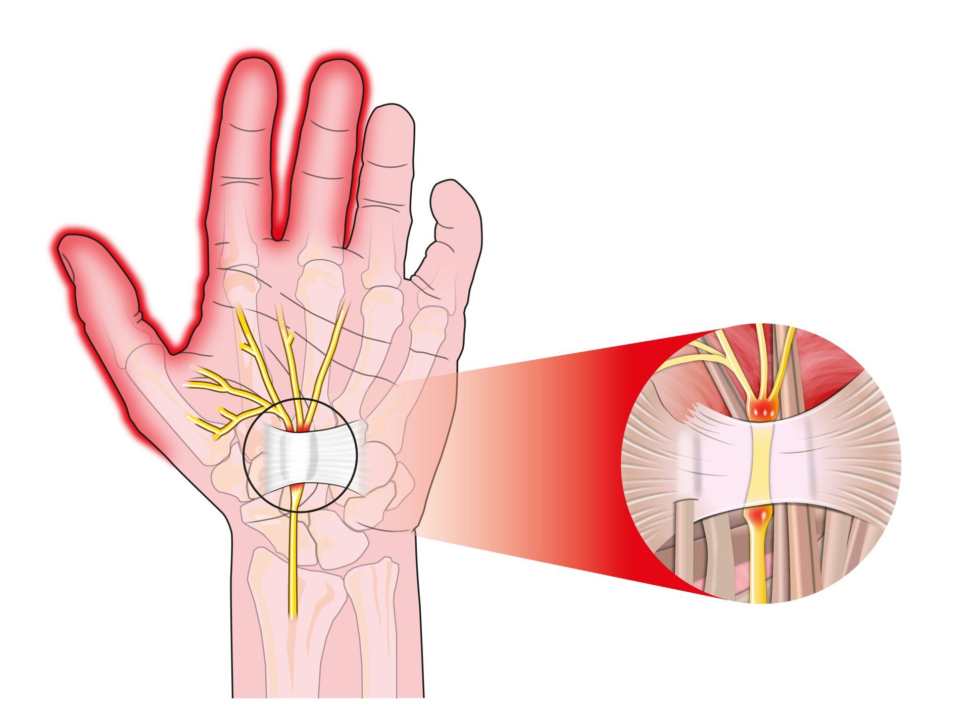
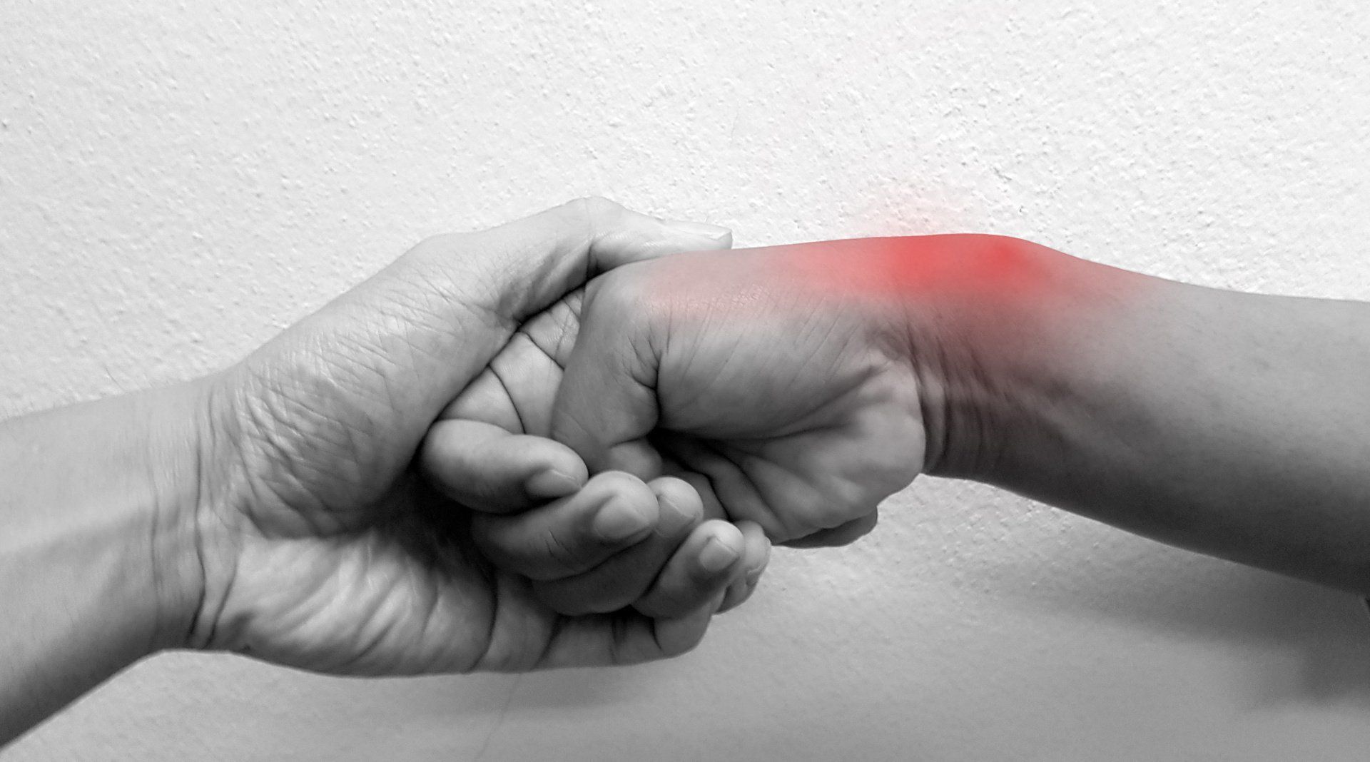
Introduction Carpal tunnel syndrome is a common condition that causes numbness, tingling and weakness in the hand specifically affecting the thumb, index and middle fingers: The little and ring fingers are not affected as they are supplied by another nerve called the ulnar nerve It is the commonest cause of peripheral nerve entrapment It is caused by compression of the median nerve as it passes from the forearm into the hand through a passage called carpal (i.e. wrist) tunnel The median nerve is one of three main nerves that supply the upper limb: The other two nerves are the ulnar nerve and the radial nerve
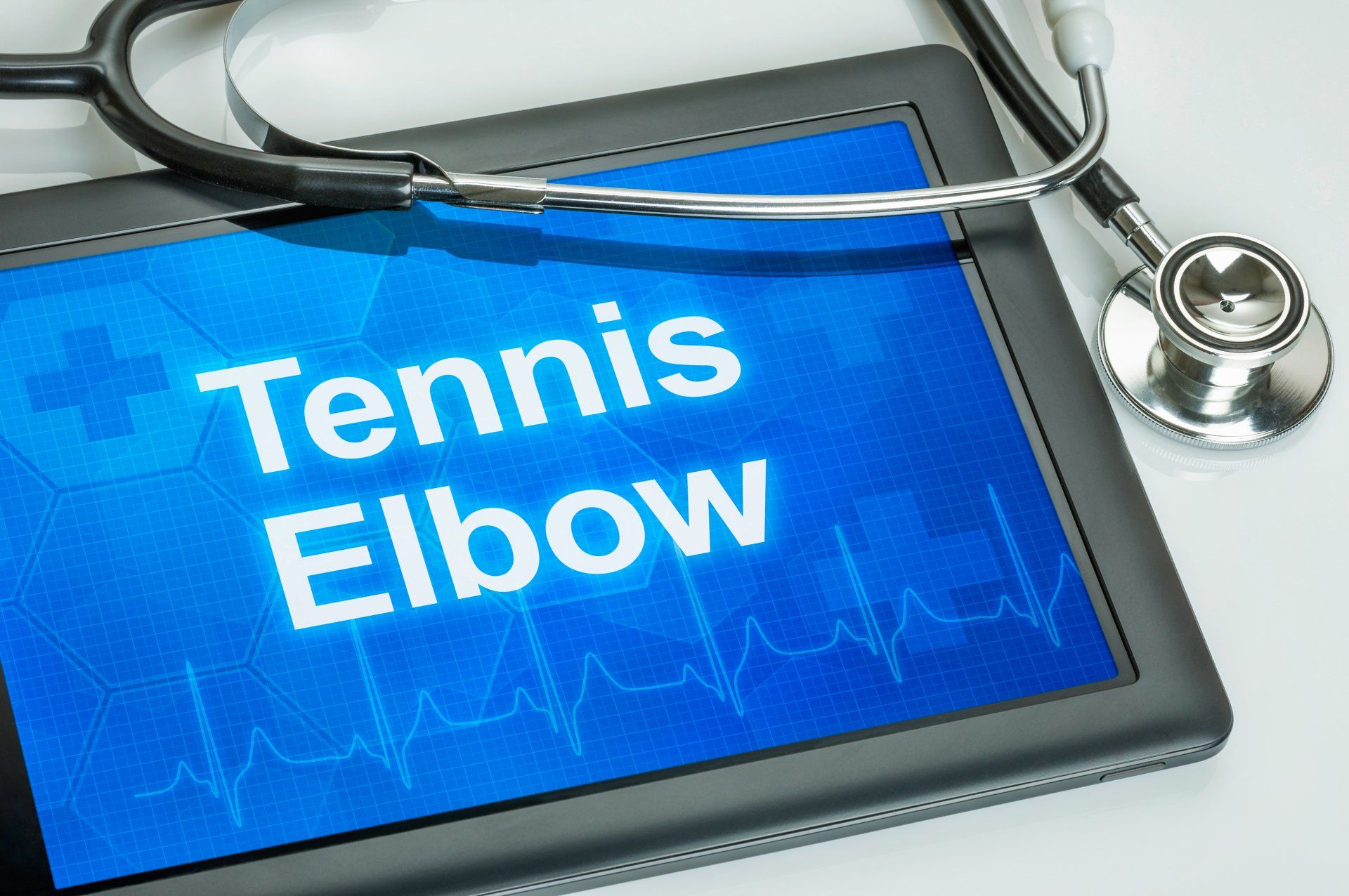
Introduction Tennis elbow (also known as lateral epicondylitis) is an overuse injury of the forearm tendons that originate over the lateral epicondyle of the humerus (bony prominence on the outside of the elbow) and act to bring the wrist backward away from the palm Whilst tennis players are particular prone to this condition it does not occur exclusively to them

Introduction This term is also known as repetitive motion or stress injury and occurs as a result of carrying out the same motion repeatedly over time causing injury to muscles and tendons It is associated with repetitive tasks, sustained or awkward position, forceful exertion, vibration or compressive forces It can affect almost any joint in the body Most commonly affected areas are hands, wrists, shoulders and neck It is thought to affect 5-10% of the general population but can be as high as 20-40% in specific working populations
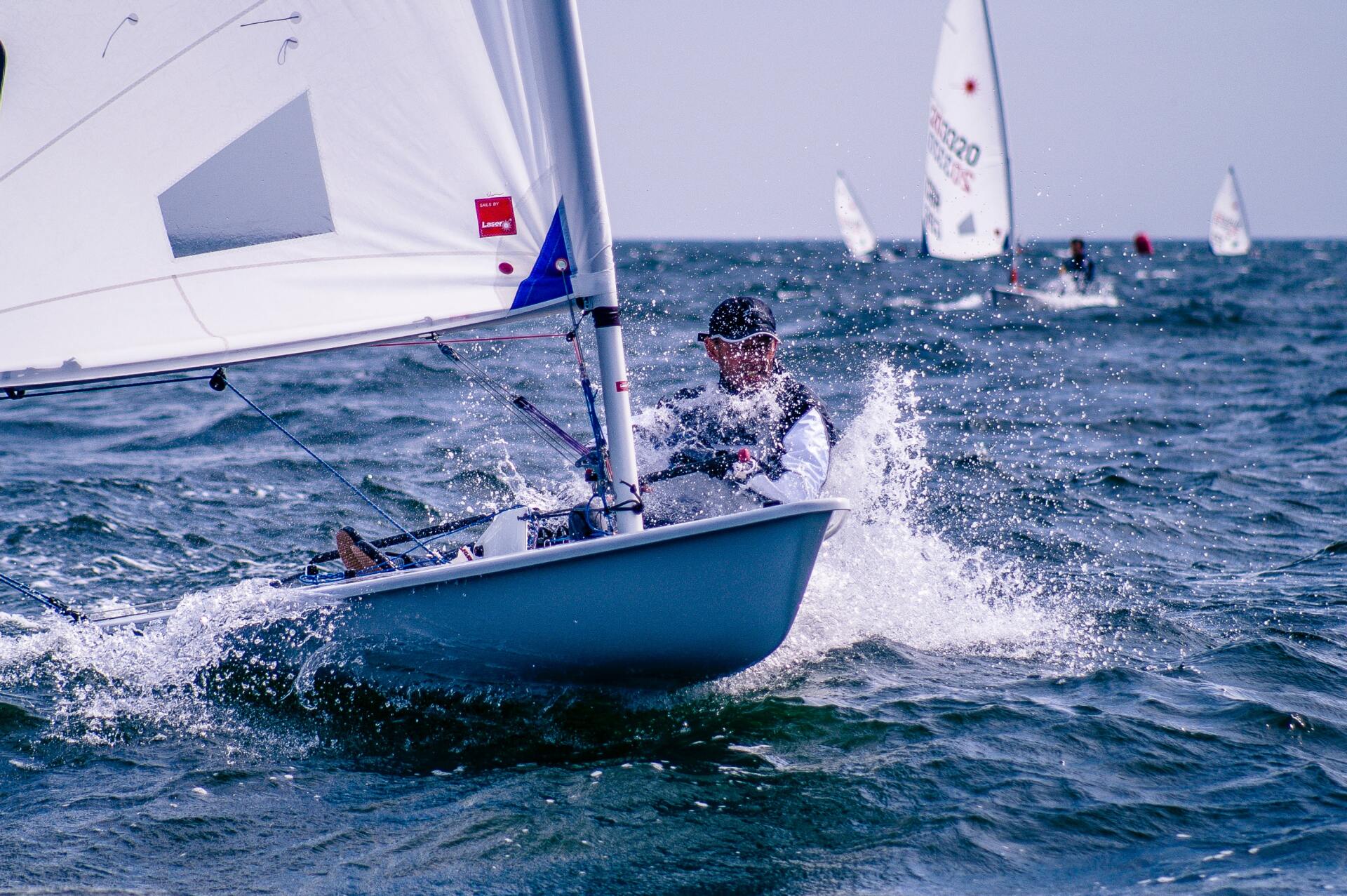
The most common injuries in sailing, are in the waist (45%), knees (30%) and shoulders (20%). Causes of sailing injuries, windsurfing injuries, sprain or ruptured knee ligament. Injury prevention, protective clothing to avoid injuries in sailing.

Injuries such as concussion, anterior cruciate ligament injury, anterior gonalgia, fatigue fracture, tibial periostitis, ankle sprain, rotator cuff injury should be avoided for women to exercise.

Women have a 50% higher risk of developing pathologies when running and exercising. Prevention of Sport Injuries.

It is composed of three components: Low energy availability with or without eating disorder - Menstrual disorders - Decreased bone mineral density (BMD) - Treatment

What is the function of vitamin D - The benefits of vitamin D, what amount of vitamin D should I take, which foods are high in vitamin D, risks of vitamin D deficiency, the role of the sun, osteoporosis, causes, symptoms.